


تاريخ الفيزياء
علماء الفيزياء


الفيزياء الكلاسيكية
الميكانيك
الديناميكا الحرارية
الكهربائية والمغناطيسية
الكهربائية
المغناطيسية
الكهرومغناطيسية
علم البصريات
تاريخ علم البصريات
الضوء
مواضيع عامة في علم البصريات
الصوت
الفيزياء الحديثة
النظرية النسبية
النظرية النسبية الخاصة
النظرية النسبية العامة
مواضيع عامة في النظرية النسبية
ميكانيكا الكم
الفيزياء الذرية
الفيزياء الجزيئية
الفيزياء النووية
مواضيع عامة في الفيزياء النووية
النشاط الاشعاعي
فيزياء الحالة الصلبة
الموصلات
أشباه الموصلات
العوازل
مواضيع عامة في الفيزياء الصلبة
فيزياء الجوامد
الليزر
أنواع الليزر
بعض تطبيقات الليزر
مواضيع عامة في الليزر
علم الفلك
تاريخ وعلماء علم الفلك
الثقوب السوداء
المجموعة الشمسية
الشمس
كوكب عطارد
كوكب الزهرة
كوكب الأرض
كوكب المريخ
كوكب المشتري
كوكب زحل
كوكب أورانوس
كوكب نبتون
كوكب بلوتو
القمر
كواكب ومواضيع اخرى
مواضيع عامة في علم الفلك
النجوم
البلازما
الألكترونيات
خواص المادة
الطاقة البديلة
الطاقة الشمسية
مواضيع عامة في الطاقة البديلة
المد والجزر
فيزياء الجسيمات
الفيزياء والعلوم الأخرى
الفيزياء الكيميائية
الفيزياء الرياضية
الفيزياء الحيوية
الفيزياء والفلسفة
الفيزياء العامة
مواضيع عامة في الفيزياء
تجارب فيزيائية
مصطلحات وتعاريف فيزيائية
وحدات القياس الفيزيائية
طرائف الفيزياء
مواضيع اخرى

Physiochemistry of color vision
 المؤلف:
Richard Feynman, Robert Leighton and Matthew Sands
المؤلف:
Richard Feynman, Robert Leighton and Matthew Sands
 المصدر:
The Feynman Lectures on Physics
المصدر:
The Feynman Lectures on Physics
 الجزء والصفحة:
Volume I, Chapter 35
الجزء والصفحة:
Volume I, Chapter 35
 2024-04-06
 2032
2032


+
-
20
Now, what about checking The spectral sensitivity curves of a normal trichromat’s receptors. against actual pigments in the eye? The pigments that can be obtained from a retina consist mainly of a pigment called visual purple. The most remarkable features of this are, first, that it is in the eye of almost every vertebrate animal, and second, that its response curve fits beautifully with the sensitivity of the eye, as seen in Fig. 35–9, in which are plotted on the same scale the absorption of visual purple and the sensitivity of the dark-adapted eye. This pigment is evidently the pigment that we see with in the dark: visual purple is the pigment for the rods, and it has nothing to do with color vision. This fact was discovered in 1877. Even today it can be said that the color pigments of the cones have never been obtained in a test tube. In 1958 it could be said that the color pigments had never been seen at all. But since that time, two of them have been detected by Rushton by a very simple and beautiful technique.
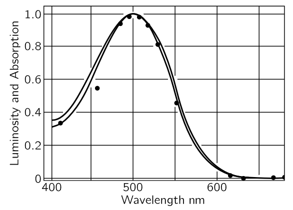
Fig. 35–9. The sensitivity curve of the dark-adapted eye, compared with the absorption curve of visual purple.
The trouble is, presumably, that since the eye is so weakly sensitive to bright light compared with light of low intensity, it needs a lot of visual purple to see with, but not much of the color pigments for seeing colors. Rushton’s idea is to leave the pigment in the eye, and measure it anyway. What he does is this. There is an instrument called an ophthalmoscope for sending light into the eye through the lens and then focusing the light that comes back out. With it one can measure how much is reflected. So, one measures the reflection coefficient of light which has gone twice through the pigment (reflected by a back layer in the eyeball, and coming out through the pigment of the cone again). Nature is not always so beautifully designed. The cones are interestingly designed so that the light that comes into the cone bounces around and works its way down into the little sensitive points at the apex. The light goes right down into the sensitive point, bounces at the bottom and comes back out again, having traversed a considerable amount of the color-vision pigment; also, by looking at the fovea, where there are no rods, one is not confused by visual purple. But the color of the retina has been seen a long time ago: it is a sort of orangey pink; then there are all the blood vessels, and the color of the material at the back, and so on. How do we know when we are looking at the pigment? Answer: First, we take a color-blind person, who has fewer pigments and for whom it is therefore easier to make the analysis. Second, the various pigments, like visual purple, have an intensity change when they are bleached by light; when we shine light on them, they change their concentration. So, while looking at the absorption spectrum of the eye, Rushton put another beam in the whole eye, which changes the concentration of the pigment, and he measured the change in the spectrum, and the difference, of course, has nothing to do with the amount of blood or the color of the reflecting layers, and so on, but only the pigment, and in this manner Rushton obtained a curve for the pigment of the protanope eye, which is given in Fig. 35–10.
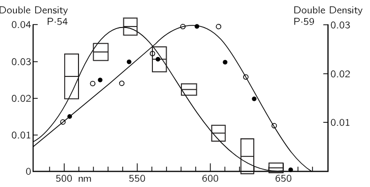
Fig. 35–10. Absorption spectrum of the color pigment of a protanope colorblind eye (squares) and a normal eye (dots).
The second curve in Fig. 35–10 is a curve obtained with a normal eye. This was obtained by taking a normal eye and, having already determined what one pigment was, bleaching the other one in the red where the first one is insensitive. Red light has no effect on the protanope eye, but does in the normal eye, and thus one can obtain the curve for the missing pigment. The shape of one curve fits beautifully with Yustova’s green curve, but the red curve is a little bit displaced. So perhaps we are getting on the right track. Or perhaps not—the latest work with deuteranopes does not show any definite pigment missing.
Color is not a question of the physics of the light itself. Color is a sensation, and the sensation for different colors is different in different circumstances. For instance, if we have a pink light, made by superimposing crossing beams of white light and red light (all we can make with white and red is pink, obviously), we may show that white light may appear blue. If we place an object in the beams, it casts two shadows—one illuminated by the white light alone and the other by the red. For most people the “white” shadow of an object looks blue, but if we keep expanding this shadow until it covers the entire screen, we see that it suddenly appears white, not blue! We can get other effects of the same nature by mixing red, yellow, and white light. Red, yellow, and white light can produce only orangey yellows, and so on. So if we mix such lights roughly equally, we get only orange light. Nevertheless, by casting different kinds of shadows in the light, with various overlaps of colors, one gets quite a series of beautiful colors which are not in the light themselves (that is only orange), but in our sensations. We clearly see many different colors that are quite unlike the “physical” ones in the beam. It is very important to appreciate that a retina is already “thinking” about the light; it is comparing what it sees in one region with what it sees in another, although not consciously.
 الاكثر قراءة في مواضيع عامة في علم البصريات
 اخر الاخبار
اخبار العتبة العباسية المقدسة

الآخبار الصحية


مواضيع ذات صلة
 قسم الشؤون الفكرية يصدر كتاباً يوثق تاريخ السدانة في العتبة العباسية المقدسة
 "المهمة".. إصدار قصصي يوثّق القصص الفائزة في مسابقة فتوى الدفاع المقدسة للقصة القصيرة
 (نوافذ).. إصدار أدبي يوثق القصص الفائزة في مسابقة الإمام العسكري (عليه السلام)


