Polysaccharides:- Bacterial and Algal Cell Walls Contain Structural Hetero polysaccharides
The rigid component of bacterial cell walls is a hetero polymer of alternating (β1 →4)-linked N-acetylglucosamine and N-acetylmuramic acid residues (Fig. 7–22). The linear polymers lie side by side in the cell wall, cross linked by short peptides, the exact structure of which depends on the bacterial species. The peptide cross-links weld the polysaccharide chains into a strong sheath that envelops the entire cell and prevents cellular swelling and lysis due to the osmotic entry of water. The enzyme lysozyme kills bacteria by hydrolyzing the (β1 →4) glycosidic bond between N-acetylglucosamine and N acetylmuramic acid (see Fig. 6–24). Lysozyme is notably present in tears, presumably as a defense against bacterial infections of the eye. It is also produced by certain bacterial viruses to ensure their release from the host bacterial cell, an essential step of the viral infection cycle. Penicillin and related antibiotics kill bacteria by preventing synthesis of the cross-links, leaving the cell wall too weak to resist osmotic lysis (see Box 20–1). Certain marine red algae, including some of the sea weeds, have cell walls that contain agar, a mixture of sulfated heteropolysaccharides made up of D-galactose and an L-galactose derivative ether-linked between C-3 and C-6 (Fig. 7–23). The two major components of agar are the unbranched polymer agarose (Mr ~120,000) and a branched component, agaropectin. The remark able gel-forming property of agarose makes it useful in the biochemistry laboratory. When a suspension of agarose in water is heated and cooled, the agarose forms a double helix: two molecules in parallel orientation twist together with a helix repeat of three residues; water molecules are trapped in the central cavity. These structures in turn associate with each other to form a gel— a three-dimensional matrix that traps large amounts of water. Agarose gels are used as inert supports for the electrophoretic separation of nucleic acids, an essential part of the DNA sequencing process (p. 8-24). Agar is also used to form a surface for the growth of bacterial colonies. Another commercial use of agar is for the capsules in which some vitamins and drugs are packaged; the dried agar material dissolves readily in the stomach and is metabolically inert.
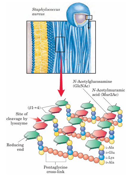
FIGURE 7–22 Peptidoglycan. Shown here is the peptidoglycan of the cell wall of Staphylococcus aureus, a gram-positive bacterium. Pep tides (strings of colored spheres) covalently link N-acetylmuramic acid residues in neighboring polysaccharide chains. Note the mixture of L and D amino acids in the peptides. Gram-positive bacteria have a pentaglycine chain in the cross-link. Gram-negative bacteria, such as E. coli, lack the pentaglycine; instead, the terminal D-Ala residue of one tetrapeptide is attached directly to a neighboring tetrapeptide through either L-Lys or a lysine-like amino acid, diaminopimelic acid.
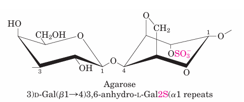
FIGURE 7–23 The structure of agarose. The repeating unit consists of D-galactose (β1 →4) -linked to 3,6-anhydro-L-galactose (in which an ether ring connects C-3 and C-6). These units are joined by (


