

النبات

مواضيع عامة في علم النبات

الجذور - السيقان - الأوراق

النباتات الوعائية واللاوعائية

البذور (مغطاة البذور - عاريات البذور)

الطحالب

النباتات الطبية


الحيوان

مواضيع عامة في علم الحيوان

علم التشريح

التنوع الإحيائي

البايلوجيا الخلوية


الأحياء المجهرية

البكتيريا

الفطريات

الطفيليات

الفايروسات


علم الأمراض

الاورام

الامراض الوراثية

الامراض المناعية

الامراض المدارية

اضطرابات الدورة الدموية

مواضيع عامة في علم الامراض

الحشرات


التقانة الإحيائية

مواضيع عامة في التقانة الإحيائية


التقنية الحيوية المكروبية

التقنية الحيوية والميكروبات

الفعاليات الحيوية

وراثة الاحياء المجهرية

تصنيف الاحياء المجهرية

الاحياء المجهرية في الطبيعة

أيض الاجهاد

التقنية الحيوية والبيئة

التقنية الحيوية والطب

التقنية الحيوية والزراعة

التقنية الحيوية والصناعة

التقنية الحيوية والطاقة

البحار والطحالب الصغيرة

عزل البروتين

هندسة الجينات


التقنية الحياتية النانوية

مفاهيم التقنية الحيوية النانوية

التراكيب النانوية والمجاهر المستخدمة في رؤيتها

تصنيع وتخليق المواد النانوية

تطبيقات التقنية النانوية والحيوية النانوية

الرقائق والمتحسسات الحيوية

المصفوفات المجهرية وحاسوب الدنا

اللقاحات

البيئة والتلوث


علم الأجنة

اعضاء التكاثر وتشكل الاعراس

الاخصاب

التشطر

العصيبة وتشكل الجسيدات

تشكل اللواحق الجنينية

تكون المعيدة وظهور الطبقات الجنينية

مقدمة لعلم الاجنة


الأحياء الجزيئي

مواضيع عامة في الاحياء الجزيئي


علم وظائف الأعضاء


الغدد

مواضيع عامة في الغدد

الغدد الصم و هرموناتها

الجسم تحت السريري

الغدة النخامية

الغدة الكظرية

الغدة التناسلية

الغدة الدرقية والجار الدرقية

الغدة البنكرياسية

الغدة الصنوبرية

مواضيع عامة في علم وظائف الاعضاء

الخلية الحيوانية

الجهاز العصبي

أعضاء الحس

الجهاز العضلي

السوائل الجسمية

الجهاز الدوري والليمف

الجهاز التنفسي

الجهاز الهضمي

الجهاز البولي


المضادات الميكروبية

مواضيع عامة في المضادات الميكروبية

مضادات البكتيريا

مضادات الفطريات

مضادات الطفيليات

مضادات الفايروسات

علم الخلية

الوراثة

الأحياء العامة

المناعة

التحليلات المرضية

الكيمياء الحيوية

مواضيع متنوعة أخرى

الانزيمات
Muscular System
المؤلف:
Kelly M. Harrell and Ronald Dudek
المصدر:
Lippincott Illustrated Reviews: Anatomy
الجزء والصفحة:
9-7-2021
3433
Muscular System
Muscles of the human body comprise three types: cardiac, smooth, and skeletal(Fig. 1 ). Cardiac muscle forms the walls of the heart. Cardiac muscle cells are striated but smaller than skeletal muscle cells and have a single nucleus per cell. Smooth muscle is primarily found in the walls of hallow viscera and blood vessels. Smooth muscle cells lack striations, have one nucleus per cell, and are small and narrow in appearance.
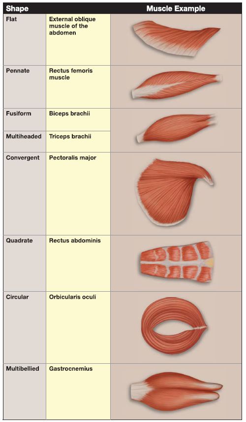
The gross Skeletal muscle cells are striated and converge to form skeletal muscles of varied shapes and sizes (Fig 2). As shown in Table 1., skeletal muscles can be described according to their shape. A skeletal muscle (e.g., biceps brachii muscle) consists of numerous fascicles, which consist of numerous skeletal muscle cells (also called skeletal muscle fibers). A skeletal muscle cell, in turn, consists of numerous myofibrils comprising thick and thin myofilaments (Fig. 3).
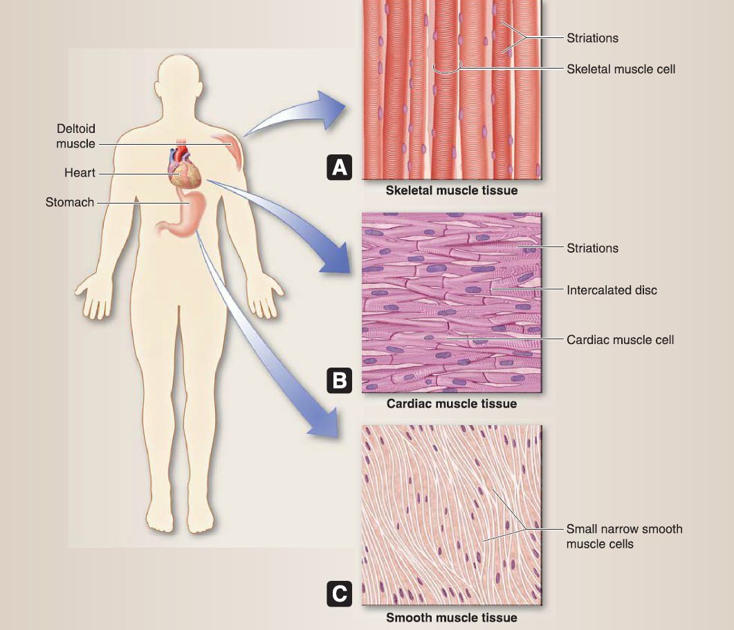
Figure 1: Muscle tissue. A, Skeletal muscle. B, Cardiac muscle. C, Smooth muscle.
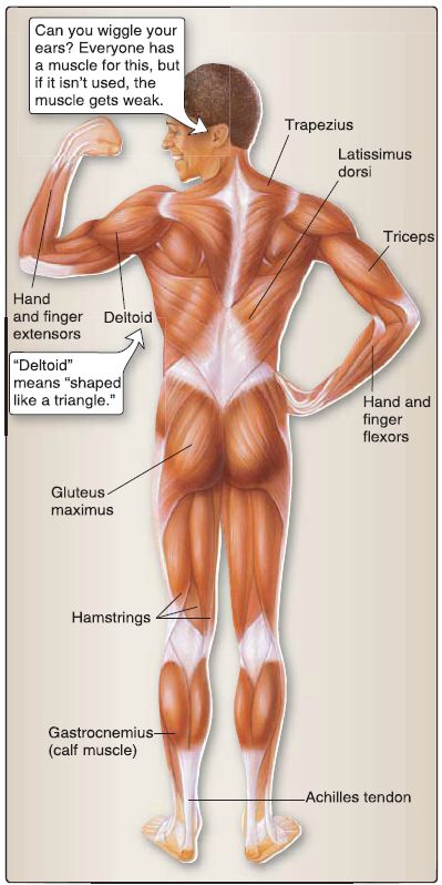
Figure 2: Skeletal muscles.
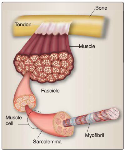
Figure 3 : Skeletal muscle organization.
Table 1: Muscle Shapes
A. Connective tissue components
The connective tissue components of skeletal muscle transmit the contractile force generated by the skeletal muscle cell to the tendon and bone so that movement of the joint occurs. The connective tissue components include the epimysium, a dense irregular connective tissue that surrounds the entire skeletal muscle; the perimysium, a dense irregular connective tissue that surrounds a bundle of skeletal muscle cells (fascicle); and the endomysium, a delicate loose connective tissue that surrounds an individual skeletal muscle cell (see Fig. 3).
B. Skeletal muscle cell
The skeletal muscle cell is cylinder shaped with tapered ends and is ~2-100 mm in length and 10-100 μm in diameter. It is multinucleated with thin, flat nuclei located at the periphery of the cell. As shown in Figure 4, its cytoplasm is characterized by striations that consist of the A band (dark), I band (light), and the Z disc. The three types of skeletal muscle cells include type I (red), type Ila (intermediate), and type llb (white). High-endurance athletes (e.g., marathon runners) have a high percentage of type I skeletal muscle cells, and low-endurance athletes (e.g., sprinters, weightlifters) have a high percentage of type llb skeletal muscle cells. Type Ila have characteristic of both types I and llb skeletal muscle cells.
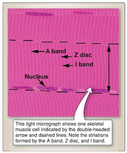
Figure 4: Skeletal muscle cell.
C. Neuromuscular junction
The neuromuscular junction is the junctional relationship of an a-motoneuron and a skeletal muscle cell in which the a-motoneuron transmits a signal to the skeletal muscle cell, thereby causing contraction of the muscle (Fig. 5). The axons of a-motoneurons whose cell bodies are located in the ventral horn of the spinal cord innervate skeletal muscle cells. The axons end as synaptic terminals with synaptic vesicles that contain the neurotransmitter acetylcholine (ACh). ACh binds to the nicotinic acetylcholine receptor (nAChR), which is a transmitter-gated ion channel located on the skeletal muscle cell. When ACh binds to nAChR, a "gate" opens and allows Na+ influx into the skeletal muscle cell, causing depolarization.
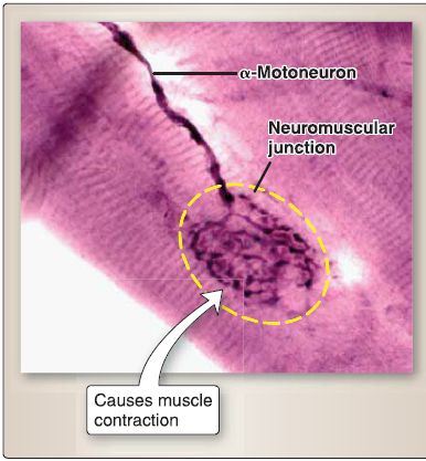
Figure 5 : Neuromuscular junction.
D. Motor unit
A single axon of an a-motoneuron may innervate 1 to 5 skeletal muscle cells, which forms a small motor unit. Or, a single axon of an a-motoneuron may branch and innervate more than 150 skeletal muscle cells, forming a large motor unit.
A motor unit is the functional contractile unit of a gross skeletal muscle, not a skeletal muscle cell.
E. Muscle spindle
As shown in Figure 6, the muscle spindle is a small, elongated, encapsulated structure distributed throughout a gross skeletal muscle that senses both dynamic changes in muscle length and static muscle length as well as activating the myotactic (stretch) reflex (e.g., knee jerk reflex). It consists of nuclear bag cells and nuclear chain cells and is innervated by type la sensory neurons (annulospiral endings), type II sensory neurons (flower-spray endings), and y-motoneurons.
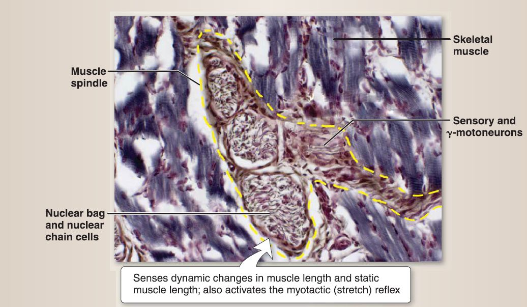 Figure 6: Muscle spindle (yellow dashed line).
 الاكثر قراءة في علم التشريح
 اخر الاخبار
اخبار العتبة العباسية المقدسة

الآخبار الصحية


 قسم الشؤون الفكرية يصدر كتاباً يوثق تاريخ السدانة في العتبة العباسية المقدسة
 "المهمة".. إصدار قصصي يوثّق القصص الفائزة في مسابقة فتوى الدفاع المقدسة للقصة القصيرة
 (نوافذ).. إصدار أدبي يوثق القصص الفائزة في مسابقة الإمام العسكري (عليه السلام)


