General considerations and theoretical background
As noted in the introduction to this chapter, many of the comments applied to spectro photometry measured in the UV-visible region also apply to fluorescence spectrophotometry. However, there are some fundamental differences which apply to fluorescence spectrophotometry; not least the molecular basis of fluorescence itself. The consequences of this are that there are some differences in instrumentation, sample treatment and data interpretation that merit a discussion of fluorescence spectrophotometry in addition to UV-visible. Molecular fluorescence is an emission process in which molecules are excited by the absorption of electromagnetic radiation. The excited species then relax to the ground state, giving up their excess energy as photons. There are several ways an excited molecule can give up its excess energy and relax to its ground state. Two of the most important of these mechanisms are nonradiative relaxation and fluorescent relaxation.
Nonradiative relaxation can occur through collisions between excited molecules and molecules of the solvent, by giving excess energy to solvent molecules. When relaxation takes place by fluorescence, bands of radiation are produced as the excited molecules relax to several energy states, which are very close in energy level and thus in wavelength (Fig. 15.13). Fluorescence occurs only from the lowest vibrational level of an excited electronic state.
Molecular fluorescence bands are made up largely of lines that are longer in wavelength (lower in energy) than the band of absorbed radiation responsible for their excitation. This shift to longer wavelength is sometimes called the Stokes’ shift. For that reason, the absorption or excitation spectrum and the fluorescence spec trum for a compound often appear as approximate mirror images of one another. The most useful region for the fluorescence technique is 200–800 nm.
Fluorescence spectrophotometry is usually the method of choice for quantitative analytical purposes if applicable. It has assumed a major role in analysis, particularly the determination of trace contaminants in our environment, industries and bodies, because for applicable compounds fluorescence spectrometry gives high sensitivity and high specificity. The selectivity of fluorescence methods is greater than that of absorption methods, as fewer substances fluoresce than absorb radiation in the UV or visible region. Furthermore, fluorescence is more selective because both the emission and the absorption spectra can be obtained. Fluorescence is usually also more sensitive than absorption methods, as it is always easier to measure a small signal against a very small zero background than to measure a small difference between large signals. However, the phenomenon of fluorescence itself is subject to more rigorous constraints on molecular structure than is absorption, such that only a relatively limited number of substances exhibit fluorescence. It is possible to introduce a fluorophore by chemical reaction (derivatisation) and this is widely employed in spectrophotometry to take advantage of the sensitivity and specificity of fluorescence.
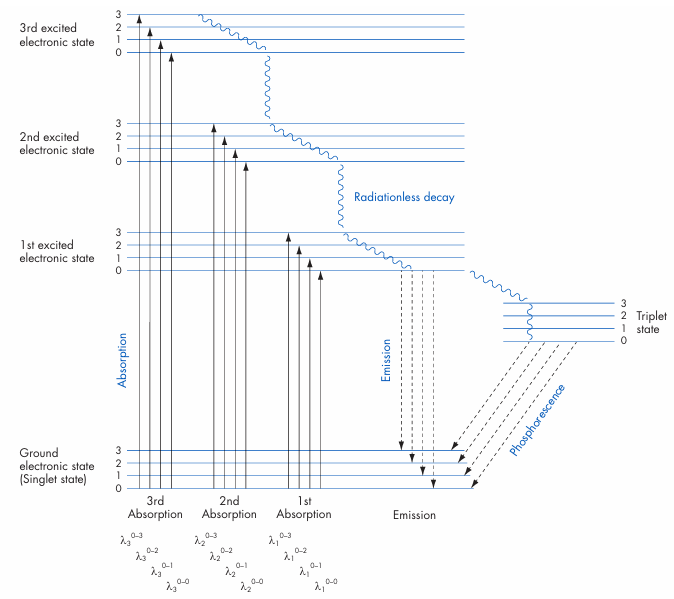
Figure 1 Normal absorption (excitation), radiationless decay, and fluorescence of an isolated chromophore.








