آخر المواضيع المضافة

 النبات
الحيوان
الأحياء المجهرية
علم الأمراض
التقانة الإحيائية
التقنية الحيوية المكروبية
التقنية الحياتية النانوية
علم الأجنة
الأحياء الجزيئي
علم وظائف الأعضاء
الغدد
المضادات الحيوية
النبات
الحيوان
الأحياء المجهرية
علم الأمراض
التقانة الإحيائية
التقنية الحيوية المكروبية
التقنية الحياتية النانوية
علم الأجنة
الأحياء الجزيئي
علم وظائف الأعضاء
الغدد
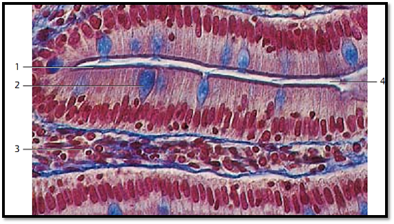
المضادات الحيوية| Microvilli, Brush Border-Duodenum |


|
|
Read More
Date: 26-7-2016
Date: 27-7-2016
Date: 18-1-2017
|
Microvilli, Brush Border-Duodenum
The cell surfaces of resorptive cells feature a dense cover of microvilli. The microvilli extend upward from the surface and create a pattern, which can be recognize d in light microscopy as a light, stripe d border, the brush border 1 . It is PAS-positive and contains several marker enzymes, which partake in resorptive cell activities. The figure shows Pseudostratified columnar epithelial surface cells from a small intestinal crypt. Goblet cells 2 , filled with secretor y products, exist side by side with the epithelial cells.
1 Brush border
2 Goblet cell with secretor y product
3 Lamina propria mucosae
4 Intestinal crypt
Stain: azan; magnification: × 160
References
Kuehnel, W.(2003). Color Atlas of Cytology, Histology, and Microscopic Anatomy. 4th edition . Institute of Anatomy Universitätzu Luebeck Luebeck, Germany . Thieme Stuttgar t · New York .

|
|
|
|
حقن الذهب في العين.. تقنية جديدة للحفاظ على البصر ؟!
|
|
|
|
|
|
|
"عراب الذكاء الاصطناعي" يثير القلق برؤيته حول سيطرة التكنولوجيا على البشرية ؟
|
|
|
|
|
|
|
جمعية العميد تعقد اجتماعها الأسبوعي لمناقشة مشاريعها البحثية والعلمية المستقبلية
|
|
|