آخر المواضيع المضافة

 النبات
الحيوان
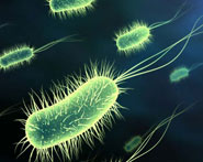
الأحياء المجهرية
علم الأمراض
التقانة الإحيائية
التقنية الحيوية المكروبية
التقنية الحياتية النانوية
علم الأجنة
الأحياء الجزيئي
علم وظائف الأعضاء
الغدد
المضادات الحيوية
النبات
الحيوان
الأحياء المجهرية
علم الأمراض
التقانة الإحيائية
التقنية الحيوية المكروبية
التقنية الحياتية النانوية
علم الأجنة
الأحياء الجزيئي
علم وظائف الأعضاء
الغدد
المضادات الحيوية| Splenic Artery and Splenic Vein |


|
|
Read More
Date: 28-7-2016
Date: 26-7-2016
Date: 14-8-2016
|
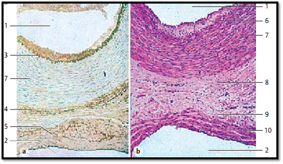
Splenic Artery and Splenic Vein
The artery is depicted in the upper part, the vein in the lower part of the photo in both figures. The three-layered structure is quite apparent in the arterial wall. This three-layered structure can also be recognized in the venous walls, albeit less clearly. The smooth muscle fibers in the tunica me dia of the vein 10 are bundled (b). The venous tunica me dia contains strong elastic networks, which become more prominent after staining with orcein (a). Part of the venous lumen 2 is shown at the lower e dges in both micrographs.
1 Arterial lumen
2 Venous lumen
3 Internal elastic lamina
4 Tunica externa of the artery
5 Tunica media of the vein
6 Internal tunica (intima) of the artery
7 Tunica media of the artery
8 Tunica externa of the artery
9 Tunica externa of the vein
10 Tunica media of the vein
Stain: a) alum hematoxylin-orcein, b) hemalum-eosin; magnification (a and b): × 15
References
Kuehnel, W.(2003). Color Atlas of Cytology, Histology, and Microscopic Anatomy. 4th edition . Institute of Anatomy Universitätzu Luebeck Luebeck, Germany . Thieme Stuttgart · New York .


|
|
|
|
حقن الذهب في العين.. تقنية جديدة للحفاظ على البصر ؟!
|
|
|
|
|
|
|
"عراب الذكاء الاصطناعي" يثير القلق برؤيته حول سيطرة التكنولوجيا على البشرية ؟
|
|
|
|
|
|
|
جمعية العميد تعقد اجتماعها الأسبوعي لمناقشة مشاريعها البحثية والعلمية المستقبلية
|
|
|