آخر المواضيع المضافة

 النبات
الحيوان
الأحياء المجهرية
علم الأمراض
التقانة الإحيائية
التقنية الحيوية المكروبية
التقنية الحياتية النانوية
علم الأجنة
الأحياء الجزيئي
علم وظائف الأعضاء
الغدد
المضادات الحيوية
النبات
الحيوان
الأحياء المجهرية
علم الأمراض
التقانة الإحيائية
التقنية الحيوية المكروبية
التقنية الحياتية النانوية
علم الأجنة
الأحياء الجزيئي
علم وظائف الأعضاء
الغدد
المضادات الحيوية| Gut- Associated Lymphoid Tissue (GALT)-Dome Areas |


|
|
Read More
Date: 9-1-2017
Date: 4-8-2016
Date:
|
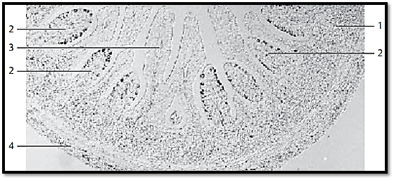
Gut- Associated Lymphoid T issue (GALT)-Dome Areas
Dome areas 2 bulge from the lymphatic tissue 1 of the submucosal tissue into the intestinal lumen. In comparison with the epithelium from mucous membranes of the large intestines 3 , the epithelium over the dome areas shows special attributes: there are no microvilli , crypts or mucous-producing goblet cells. Instead of microvilli, these specialize d epithelial cells show microfolds at their surfaces (microfolds cells, M-cells). M-cells can take up antigenic materials from the lumen and transport it to the lymphatic tissue. M-cells cannot be identified using histological routine preparations and light microscopy. M-cells from a rabbit contain a lot of vimentin as a component of intermediary filaments. It was therefore use d here as a marker for the presence of M-cells. The immunohistochemical localization of vimentin pro-duce d black precipitates in the epithelium. Vermiform appendix of a rabbit.
1 Lymph follicles
2 Dome areas
3 Mucous membrane from the large intestine
4 Tunica muscularis
Stain: immunohistochemical localization of vimentin; differential interference contrast im-aging; magnification: × 80
References
Kuehnel, W.(2003). Color Atlas of Cytology, Histology, and Microscopic Anatomy. 4th edition . Institute of Anatomy Universitätzu Luebeck Luebeck, Germany . Thieme Stuttgart · New York .

|
|
|
|
حمية العقل.. نظام صحي لإطالة شباب دماغك
|
|
|
|
|
|
|
إيرباص تكشف عن نموذج تجريبي من نصف طائرة ونصف هليكوبتر
|
|
|
|
|
|
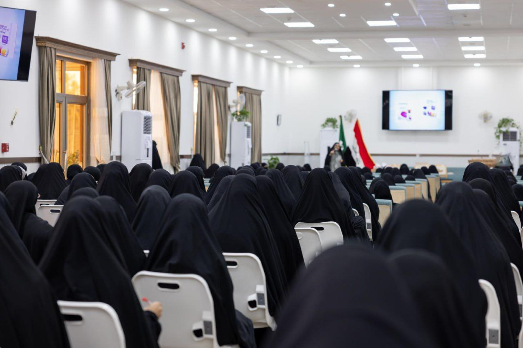
شعبة مدارس الكفيل النسوية تطلق فعّاليات مخيم (بنات العقيدة) العشرين
|
|
|
|
قسم التربية والتعليم يعقد اجتماعاً تحضيرياً لملاكاته استعداداً لانطلاق برنامجه التأهيلي
|
|
|
|
متحف الكفيل يعقد أولى اجتماعاته التحضيرية لمؤتمره الدوليّ الخامس
|
|
|
|
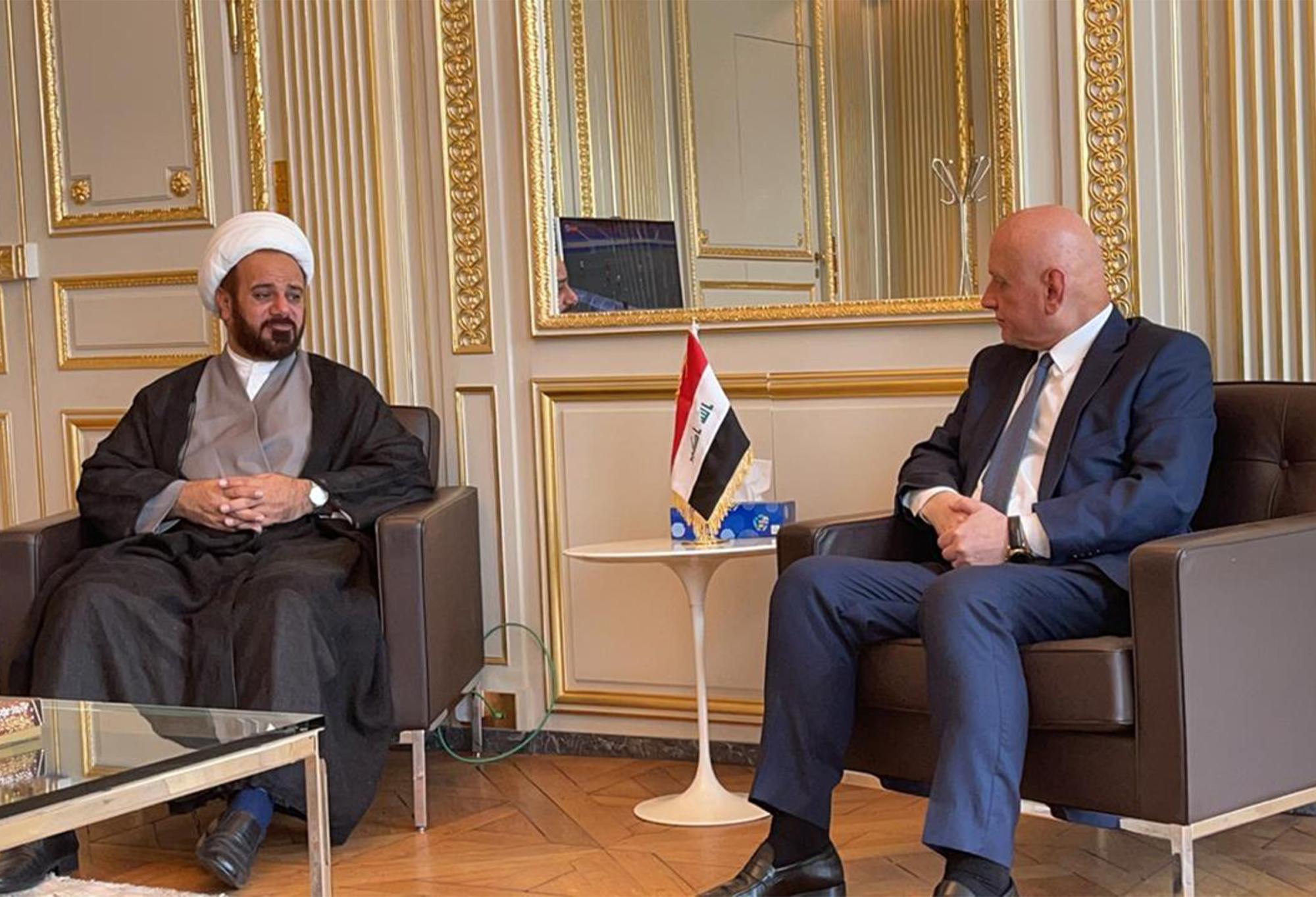
وفد قسم الشؤون الفكرية وسفير العراق في فرنسا يبحثان سبل تعزيز التعاون المشترك
|